Mesothelioma may be a neoplasm originating from serous membrane or serosa surfaces; this condition is typically related to activity exposure to amphibole. Wagner et al connected amphibole to carcinoma during a classic 1960 study of thirty three patients with carcinoma World Health Organization were exposed to amphibole during a mining space in South Africa's North Western province. [1] Of the thirty three patients, thirty two had been exposed to crocidolite, the foremost malignant neoplastic disease kind of amphibole.
Asbestos mining and production peaked from the 1930s-1960s, and amphibole was employed in a range of merchandise starting from construction provides to brake linings. throughout war II, many thousands of civilian and military staff, through their occupations, were exposed to amphibole. Production slowed dramatically within the Seventies because the health risks of amphibole became famous. Governmental restrictions were placed on its use, and various materials became accessible. Despite these changes, amphibole continues to be employed in the manufacture of some fireplace safety merchandise.
The clinical phase between amphibole exposure and carcinoma development is 35-40 years, and as a result, the quantity of carcinoma patients has continued to rise despite remittent amphibole production. the foremost common findings on physical examination (79%) area unit signs of serous membrane effusion (eg, dullness to percussion, remittent breath sounds).
The identification of carcinoma ought to be created with care. A clinical history of amphibole exposure and radiologic findings that area unit in line with carcinoma warrant inclusion of carcinoma within the medical diagnosis, however it's vital to fret that a identification of carcinoma can't be created solely with imaging studies. additional common diseases, like benign asbestos-related serous membrane malady and pathological process glandular carcinoma, will look radiographically a twin of carcinoma. diagnostic test with special staining and immunohistochemical and ultrastructural analysis area unit fully essential for the correct identification of carcinoma.
Mesothelioma is incredibly tough to treat; the treatment is typically surgical, though different treatment choices like therapy and radiation area unit used. the two primary surgical interventions area unit pleurectomy and extrapleural excision (EPP). [2]
Preferred examination
Chest radiography is that the initial screening examination, whereas X-radiation (CT) scanning is most well-liked for staging the neoplasm.
Magnetic resonance imaging (MRI) enhances CT scanning in some patients. tomography provides higher delineation of sentimental tissues (better soft-tissue contrast) and permits imaging within the mesial and lei planes.
Limitations of techniques
Chest radiography has restricted utility. The picture taking findings of carcinoma area unit nonspecific and area unit discovered in different diseases, as well as pathological process cancer, lymphoma, and benign amphibole malady. little malignant serous membrane effusions might not be discovered on customary radiographs. as an alternative, giant serous membrane effusions will obscure serous membrane thickening or masses; thus, malady extent is usually underestimated in radiographs.
CT scanning provides additional and higher data than plain radiography with relation to neoplasm characteristics and extent. though tomography is superior to CT scanning in some areas, this advantage failed to modification the operation during a study by Heelan et al. [11]
Neither CT scanning nor tomography provides associate unequivocal identification of mesothelioma; tissue diagnostic test is needed for the definitive identification.
Radiography
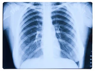
The most common carcinoma finding on radiographs is unilateral, concentric, plaquelike, or nodular serous membrane thickening (as seen within the pictures below). serous membrane effusions area unit common and should obscure the presence of the underlying serous membrane thickening. The neoplasm often extends into the fissures, that become thickened and irregular in contour. a small right-sided predominance is discovered, probably due to a bigger serous membrane extent. The neoplasm will stiffly case the respiratory organ, inflicting compression of respiratory organ parenchyma, diaphragm elevation, intercostal house narrowing, and mediastinal shift toward the neoplasm. Calcified serous membrane plaques area unit gift in 2 hundredth of patients with carcinoma and area unit sometimes associated with the previous amphibole exposure.
Lung nodules and fissure plenty sometimes result from direct carcinoma neoplasm extension into the respiratory organ parenchyma and mediastinal structures, like body fluid nodes, the serosa, and also the heart. Mechanical distortion of the hemithorax, chest wall plenty, periosteal rib reaction or rib destruction by the neoplasm area unit signs of advanced malady. though sometimes unilateral, direct extension of the neoplasm across the bodily cavity into the contralateral hemithorax will occur.
Although a certain identification can't be created on the idea of plain film findings, new unilateral serous membrane thickening or effusion during a patient World Health Organization features a history of exposure to amphibole is very implicational carcinoma.
False-positive identification supported imaging alone can be thanks to serous membrane metastases from glandular carcinoma, breast or different primary malignancies, involvement of the serosa by cancer or thymoma, or chronic infection. False-negative findings area unit potential with borderline little focus of serous membrane involvement by carcinoma.

Post a Comment for "Can a chest xray show mesothelioma?"